Е
Елена
@elena-4

Эффект slow-motion, когда Павел кружит Лику.
On-screen dialogue (characters speak with lip-sync): Лика (за кадром): "А потом было знако...

Камера внутри машины, вид с заднего сиденья. Плавное движение, показывающее их живое общение и пролетающие мимо огни города.
On-screen d...

Камера внутри машины, вид с заднего сиденья. Плавное движение, показывающее их живое общение и пролетающие мимо огни города.
On-screen d...
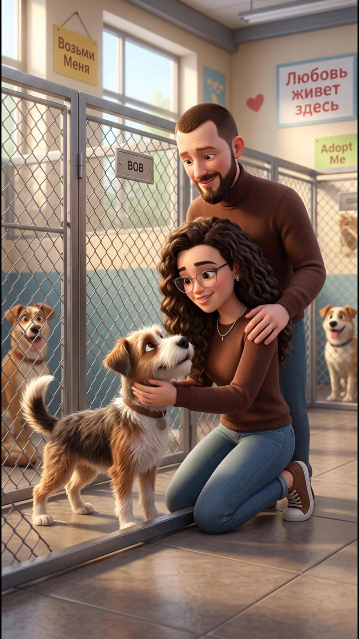
Камера немного подрагивает, создавая эффект документальной съемки. Фокус на взаимодействии Лики с животным и заботливом взгляде Павла.гол...

Камера сбоку, показывает их двоих. Плавный зум на коробку с синнабонами в момент, когда Лика ее открывает. Вокруг легкий пар от теплой вы...

Медленный облет камеры вокруг букета, фокус смещается с одного цветка на другой, подчеркивая их красоту и свежесть.
On-screen dialogue (...

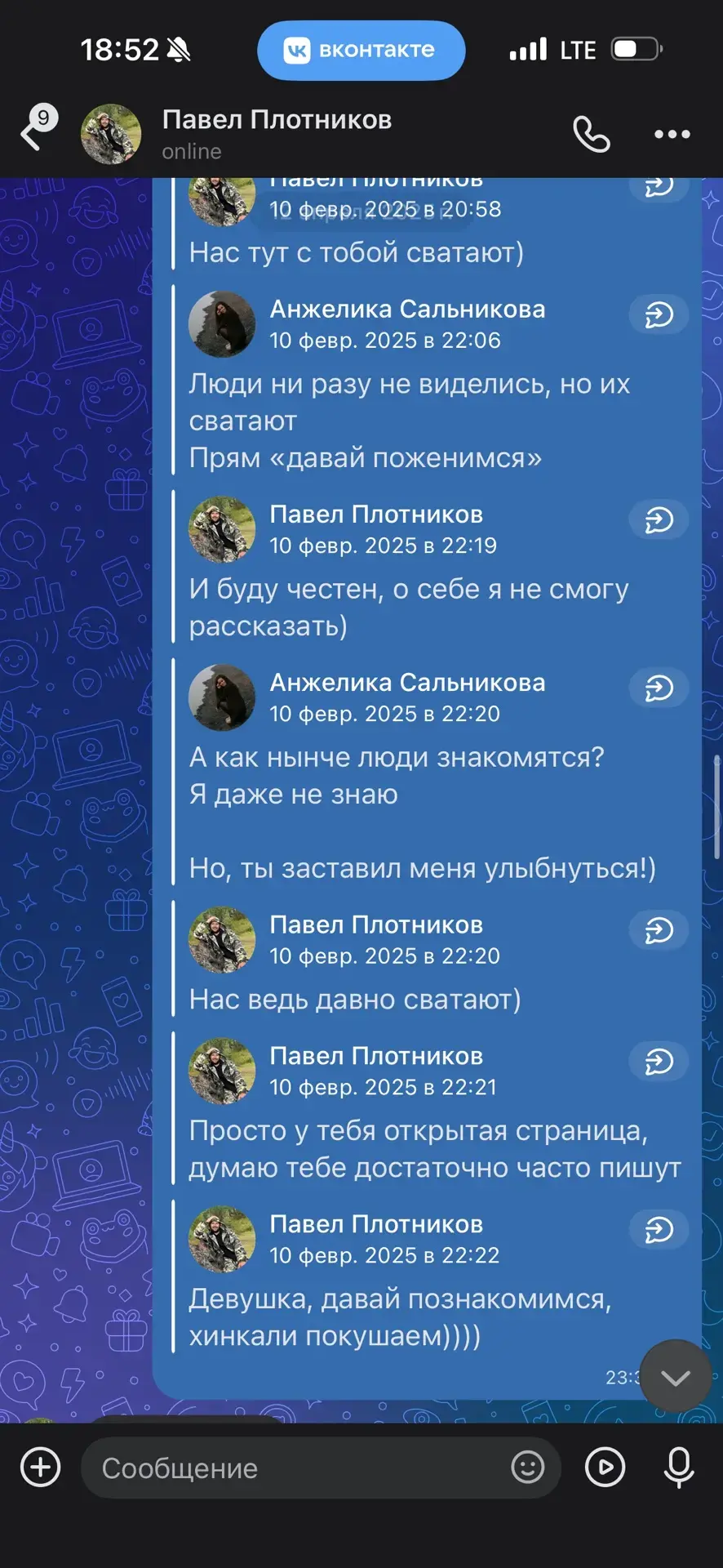
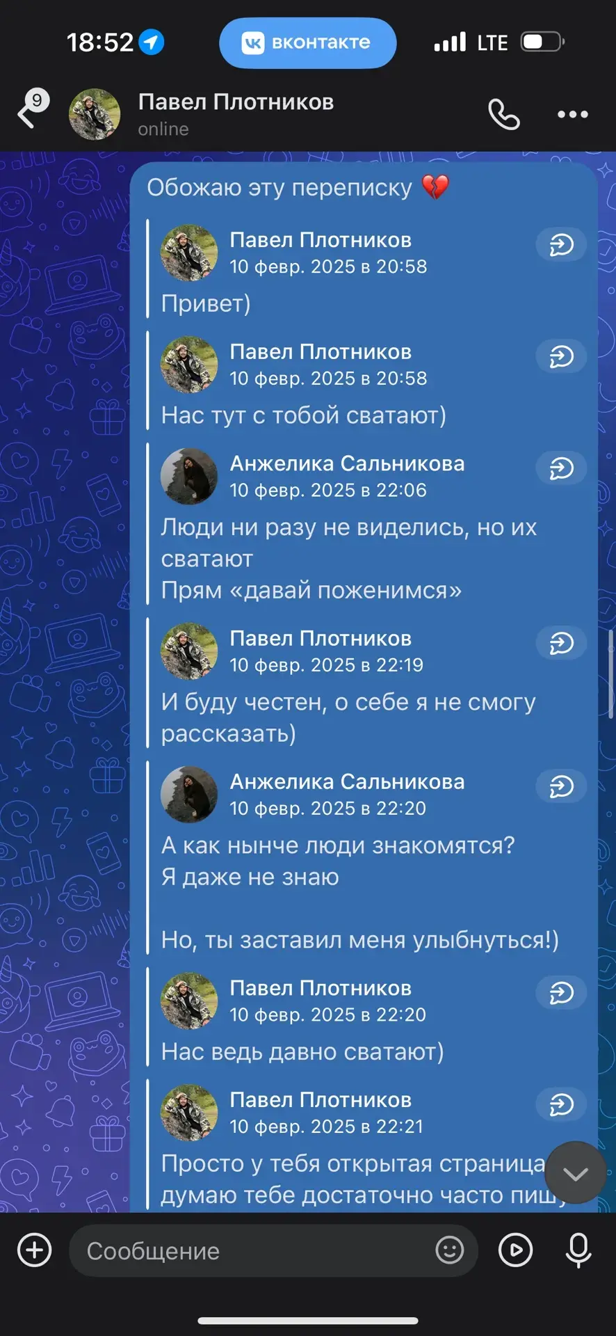
Плавный наезд на экран телефона, чтобы зритель мог прочитать текст,текст точь в точь как на картинке,ничего не менять. Легкое свечение эк...

Камера внутри машины, вид с заднего сиденья. Плавное движение, показывающее их живое общение и пролетающие мимо огни города.
On-screen d...
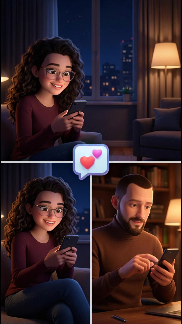
Быстрые переходы между кадрами Лики и Павла, имитирующие динамику переписки. На экране пролетают "лайки" и сердечки.
On-screen dialogue ...
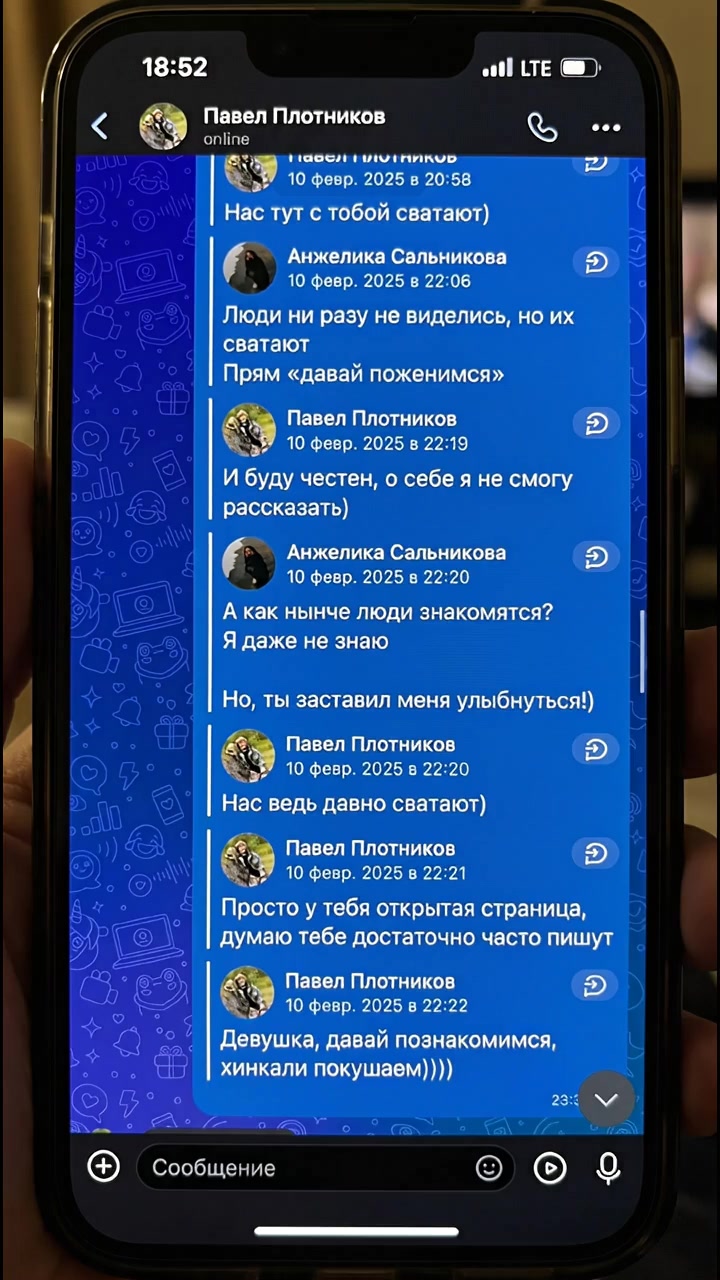
Плавный наезд на экран телефона, чтобы зритель мог прочитать текст. Легкое свечение экрана освещает фон. Видно только экран телефона и пе...

Быстрые переходы между кадрами Лики и Павла, имитирующие динамику переписки. На экране пролетают "лайки" и сердечки.
On-screen dialogue ...
Плавный наезд на экран телефона, чтобы зритель мог прочитать текст. Легкое свечение экрана освещает фон.
On-screen dialogue (characters ...
 image
image
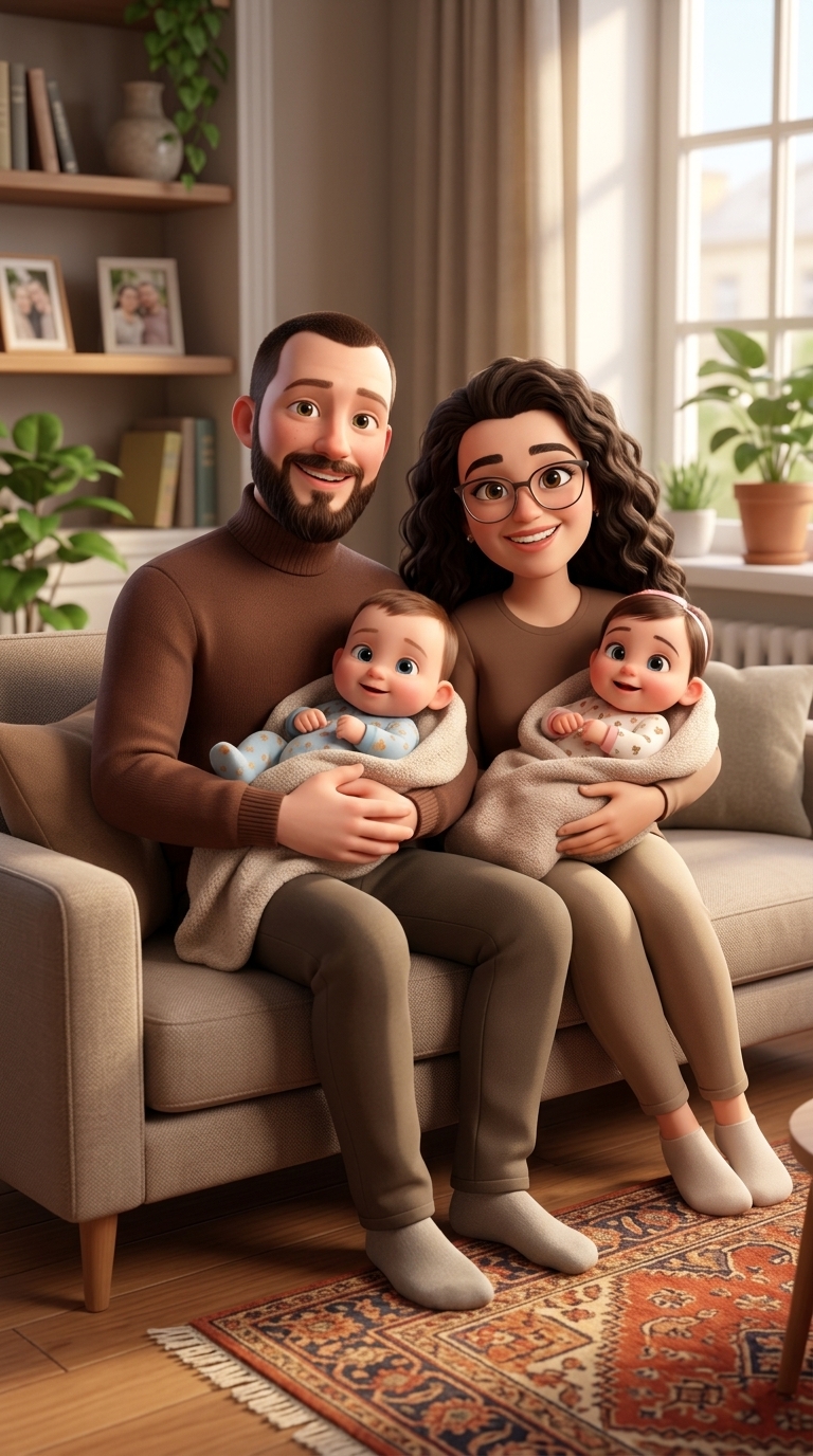
Art style: 3D Pixar.
Павел и Лика сидят на диване в современной уютной гостинной ,на руках у них маленькие дети двойняшки .
━━━━━━━━━━...
 image
image
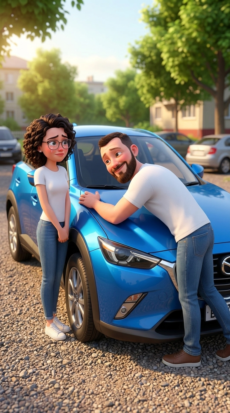
Art style: 3D Pixar.
Паша обнимает капот синего кроссовера с референта. Павел одет в белую футболку и джинцы. Лика стоит рядом с мокрыми...
 image
image
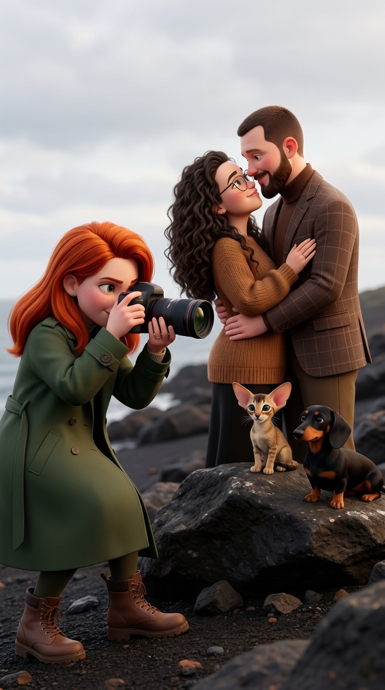
Art style: 3D Pixar.
Сцена фотосессии в стиле "лав стори". .фото с референта + Профессиональный фотограф девушка с рыжими волосами ловит...
 image
image
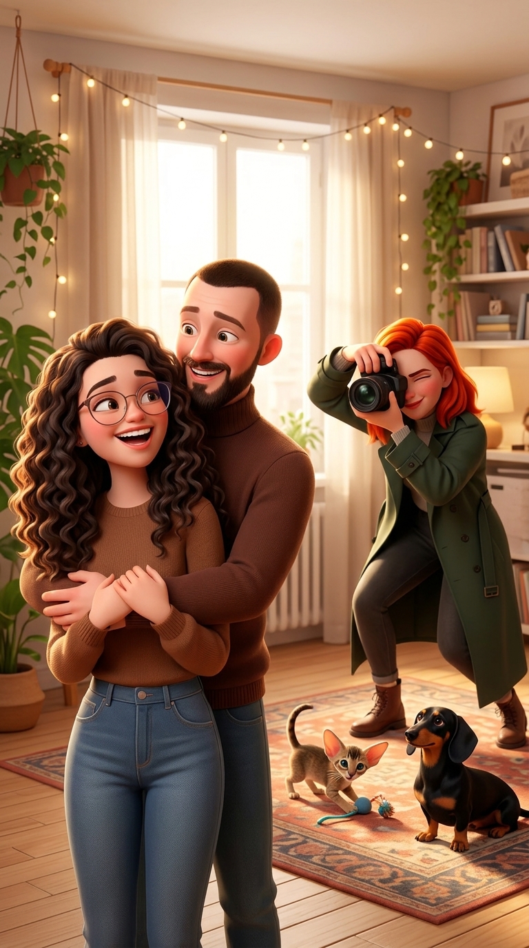
Art style: 3D Pixar.
Сцена фотосессии в стиле "лав стори". .фото с референта + Профессиональный фотограф девушка с рыжими волосами ловит...
 image
image
Art style: 3D Pixar.
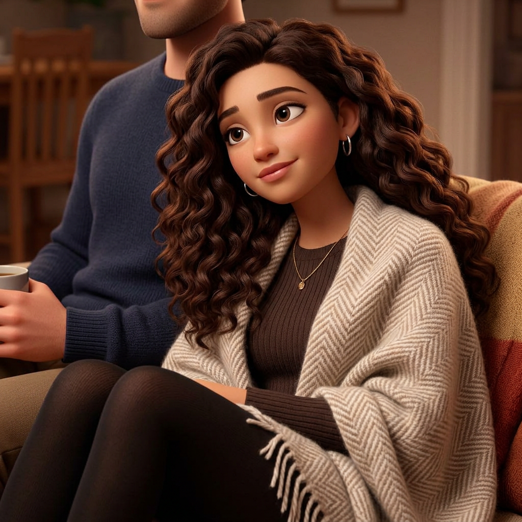
Молодая женщина лет 25 с овальным лицом и легким оливковым оттенком кожи. У нее густые, длинные, темно-каштановые в...
 image
image
Art style: 3D Pixar.
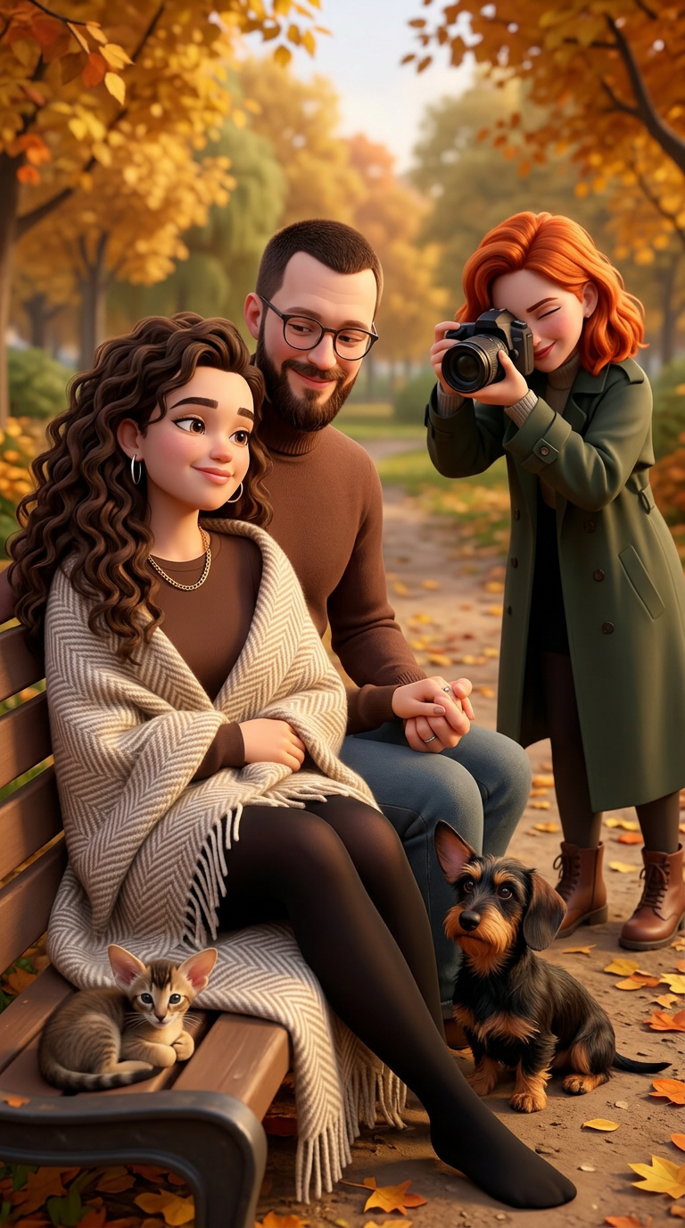
Сцена фотосессии в стиле "лав стори". .фото с референта + Профессиональный фотограф девушка с рыжими волосами ловит...
 image
image
Art style: 3D Pixar.
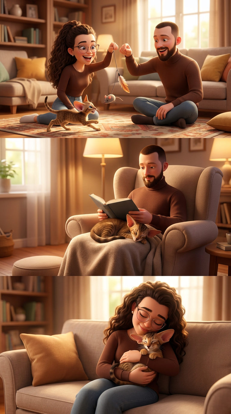
Монтаж счастливых моментов: Лика и Павел играют с котенком Бруно с референса на полу с помощью игрушечной мышки. Ко...
 image
image
Art style: 3D Pixar.
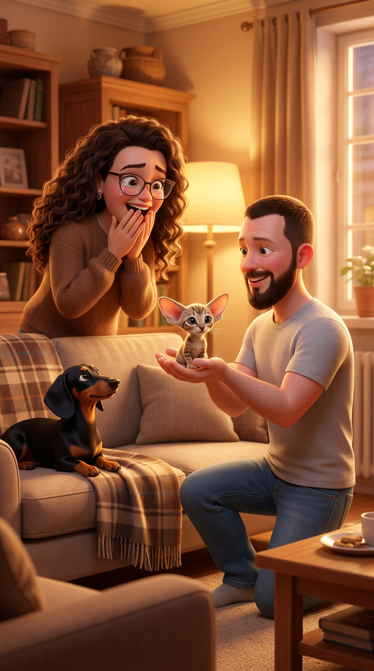
Уютная гостиная.Павел одет в светлую футболку и джинцы. Павел делает сюрприз Лике, протягивая ей маленького котенка...
 image
image
Art style: 3D Pixar.
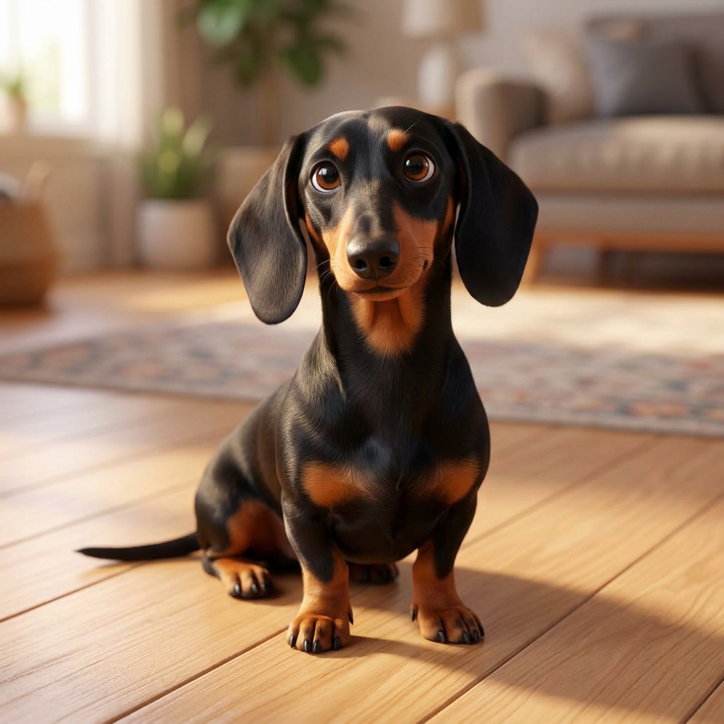
Взрослая такса с гладкой черной шерстью и рыжими подпалинами сидит на светлом полу в помещении. У собаки внимательн...
 image
image
Art style: 3D Pixar.
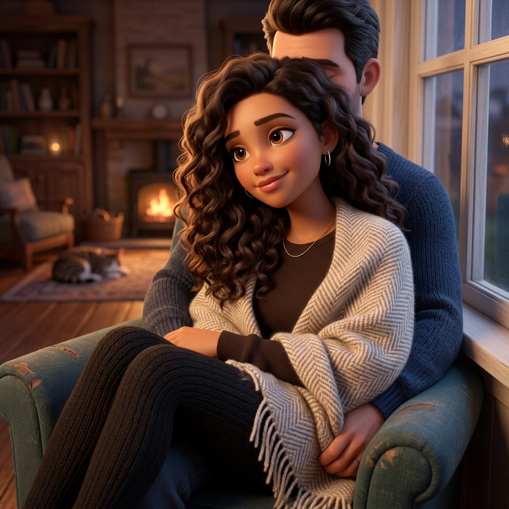
Молодая славянская женщина около 25 лет с овальным лицом, нежной светлой кожей и мягкими чертами. У неё длинные вол...
 image
image
Art style: 3D Pixar.
Молодая женщина лет 25 с овальным лицом и легким оливковым оттенком кожи. У нее густые, длинные, темно-каштановые в...
 image
image
Art style: 3D Pixar.
Мужчина около 30-35 лет в стиле 3D-анимации с овальным лицом и светлой кожей. У него короткая стрижка «ёжик» и густ...
 image
image
Art style: 3D Pixar.
Молодая женщина около 20 лет с овальным лицом и светлой кожей в стиле 3D-анимации. У нее длинные, густые, темно-каш...
 image
image
Art style: 3D Pixar.
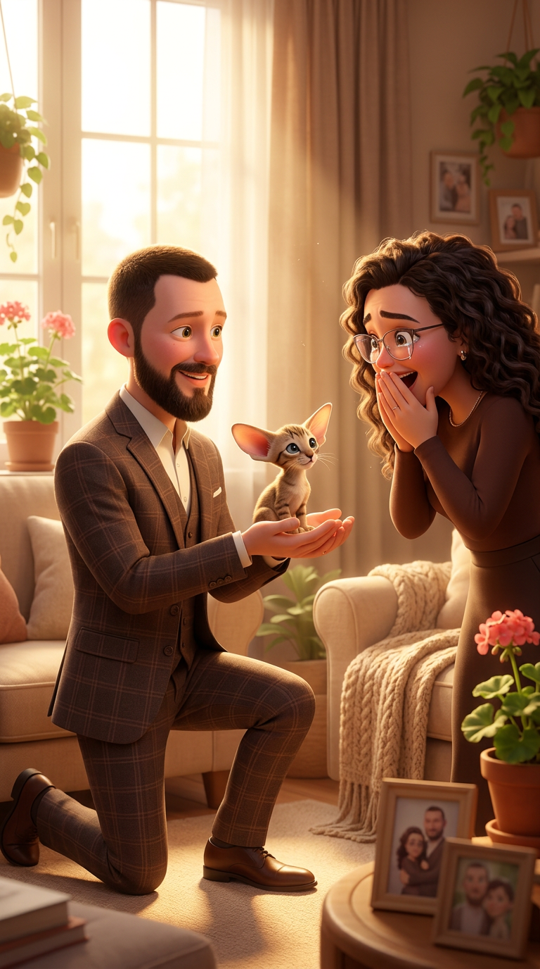
Уютная гостиная. Павел делает сюрприз Лике, протягивая ей маленького котенка Бруно. Лицо Лики выражает шок и востор...
 image
image
Art style: 3D Pixar.
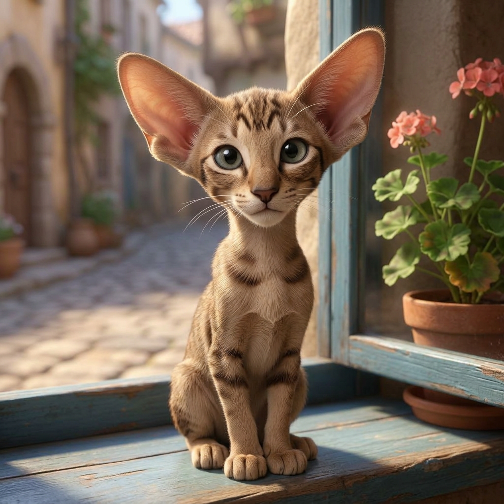
Молодой котенок ориентальной породы с длинной клиновидной мордочкой и выразительным крупным носом. У него очень кор...
 image
image
Art style: 3D Pixar.
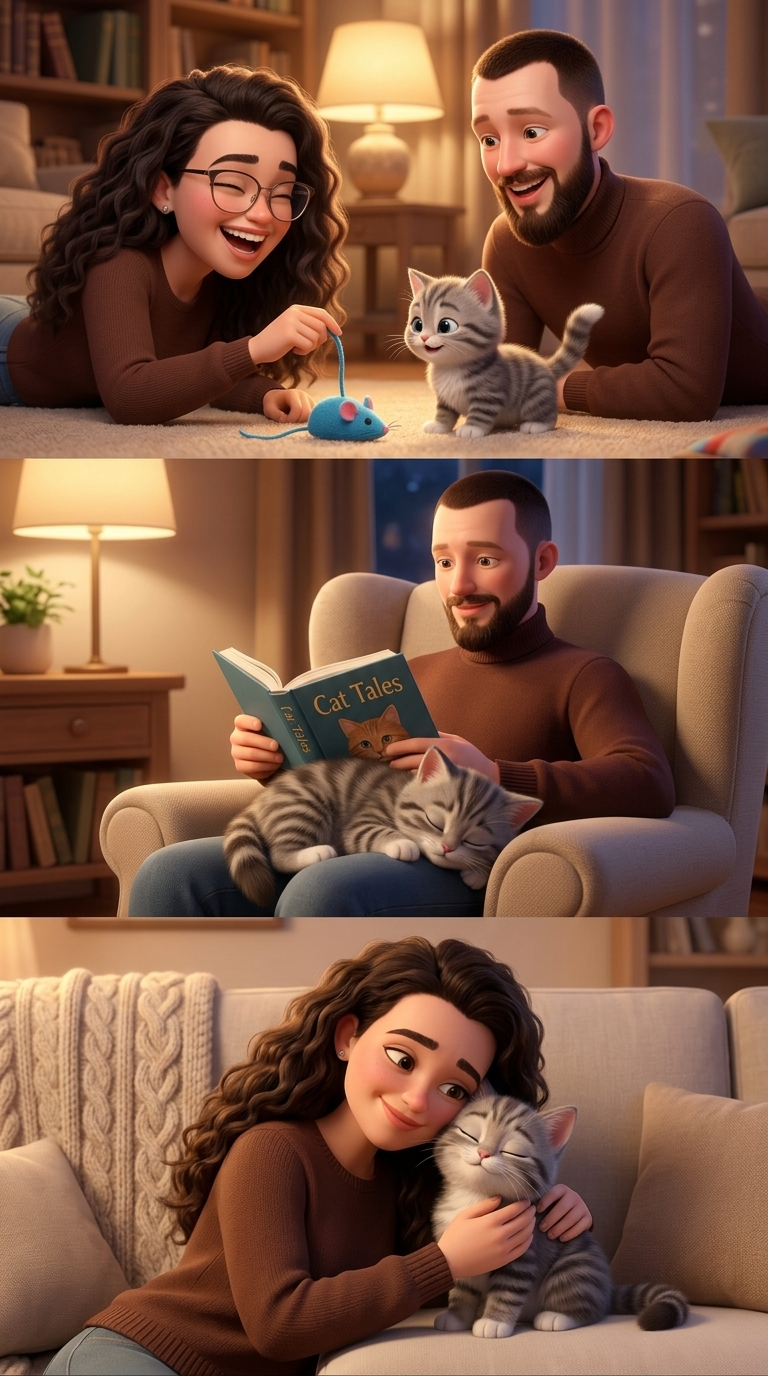
Монтаж счастливых моментов: Лика и Павел играют с котенком Бруно с референса на полу с помощью игрушечной мышки. Ко...
 image
image
Art style: 3D Pixar.
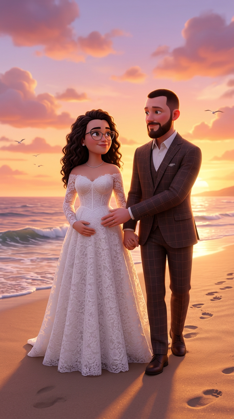
Финальная сцена. Лика и Павел гуляют по пляжу на закате, держась за руки. Павел нежно кладет руку на живот Лики. Он...
 image
image
Art style: 3D Pixar.
Финальная сцена. Лика и Павел гуляют по пляжу на закате, держась за руки. Павел нежно кладет руку на живот Лики. Он...
 image
image
Art style: 3D Pixar.
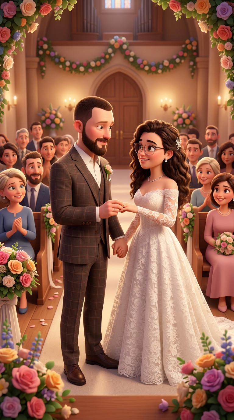
Сцена свадьбы. Павел и Лика, в свадебных нарядах, стоят у алтаря. Они смотрят друг на друга с любовью и нежностью, ...
 image
image
Art style: 3D Pixar.
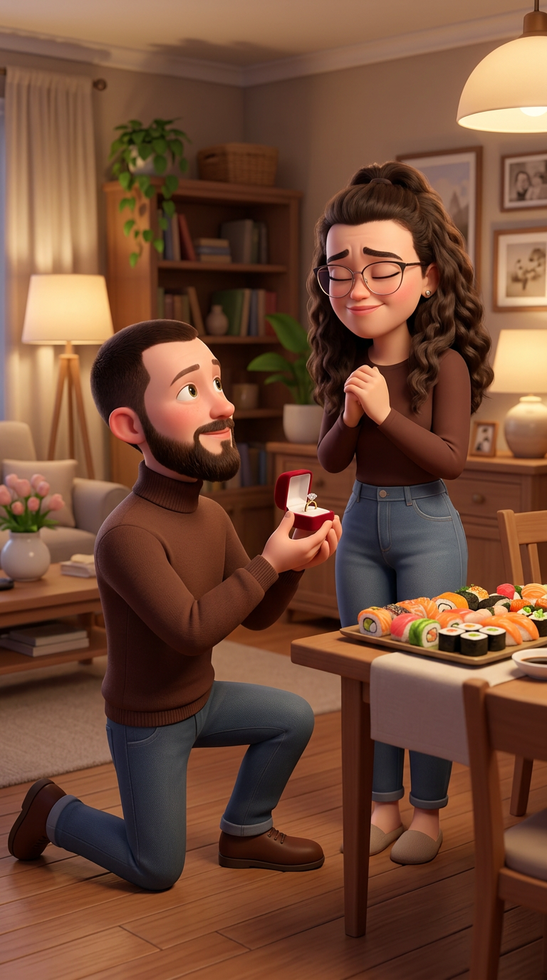
Домашняя обстановка. На столе сет из суши и роллов. Павел стоит на одном колене перед Ликой, у которой закрыты глаз...
 image
image
Art style: 3D Pixar.
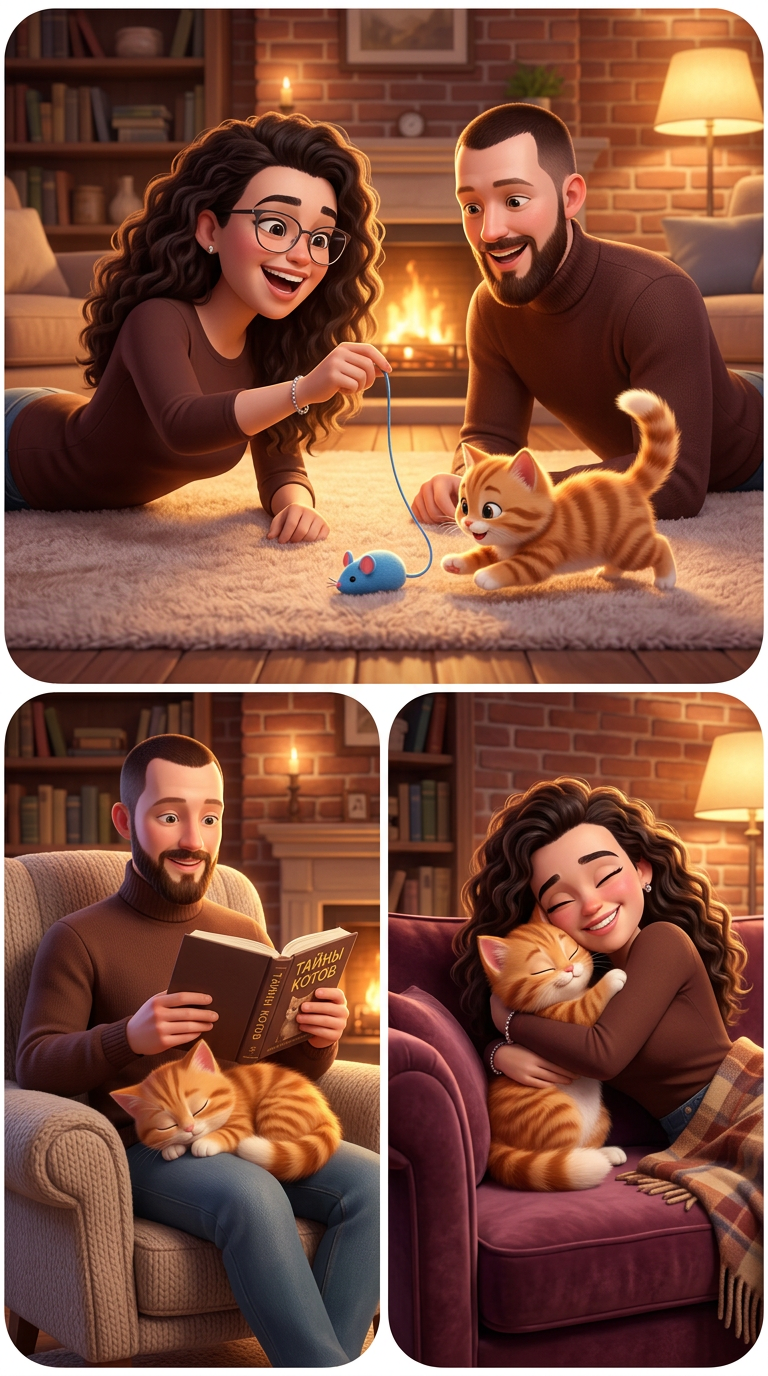
Монтаж счастливых моментов: Лика и Павел играют с котенком на полу с помощью игрушечной мышки. Котенок спит на коле...
 image
image
Art style: 3D Pixar.
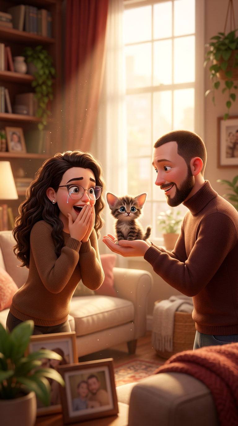
Уютная гостиная. Павел делает сюрприз Лике, протягивая ей маленького пушистого котенка. Лицо Лики выражает шок и во...
 image
image
Image - 2026-05-28 11:51
 image
image
Art style: 3D Pixar.
Величественный горный пейзаж Хакасии на закате. Павел и Лика стоят на вершине, обнявшись, и смотрят на открывающийс...
 image
image
Art style: 3D Pixar.
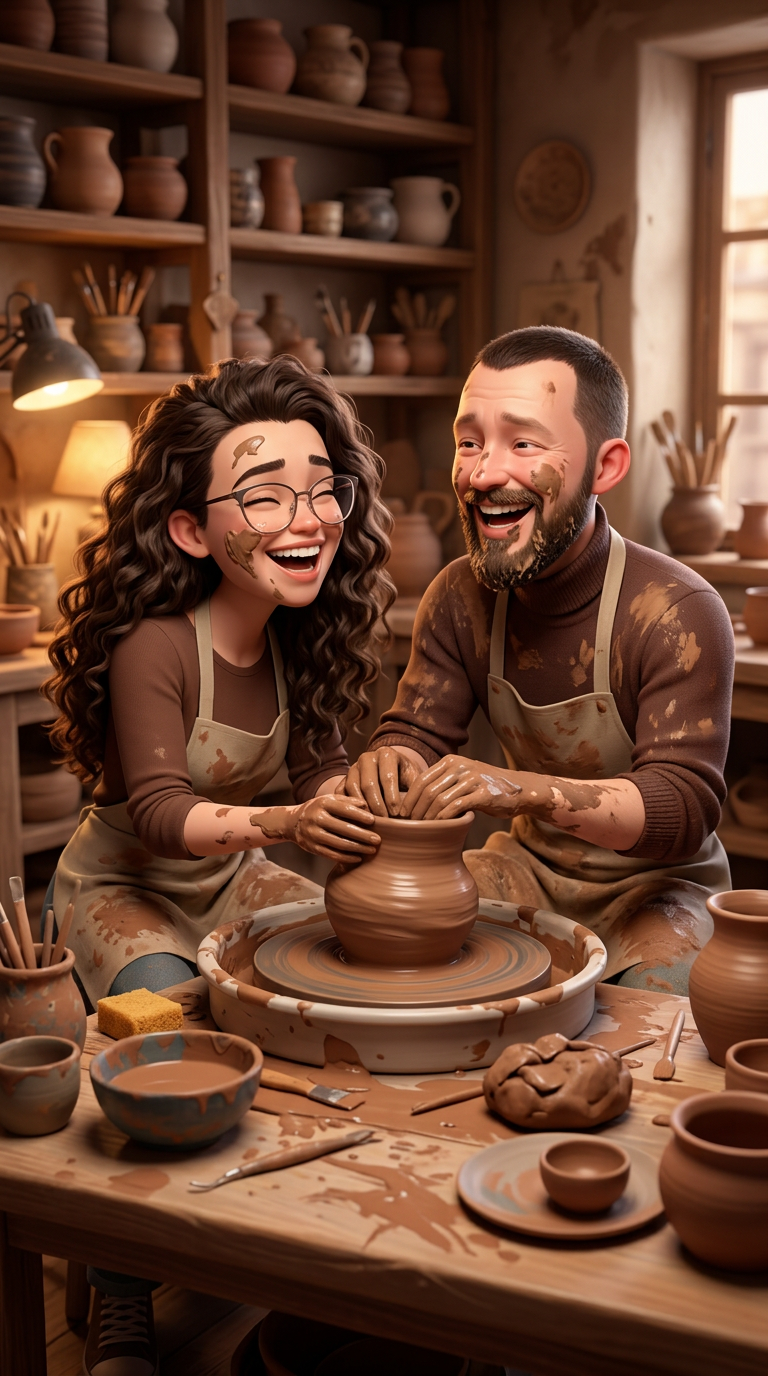
Лика и Павел сидят за гончарным кругом, их руки вместе в глине, они пытаются слепить горшок и смеются, измазавшись....
 image
image
Art style: 3D Pixar.
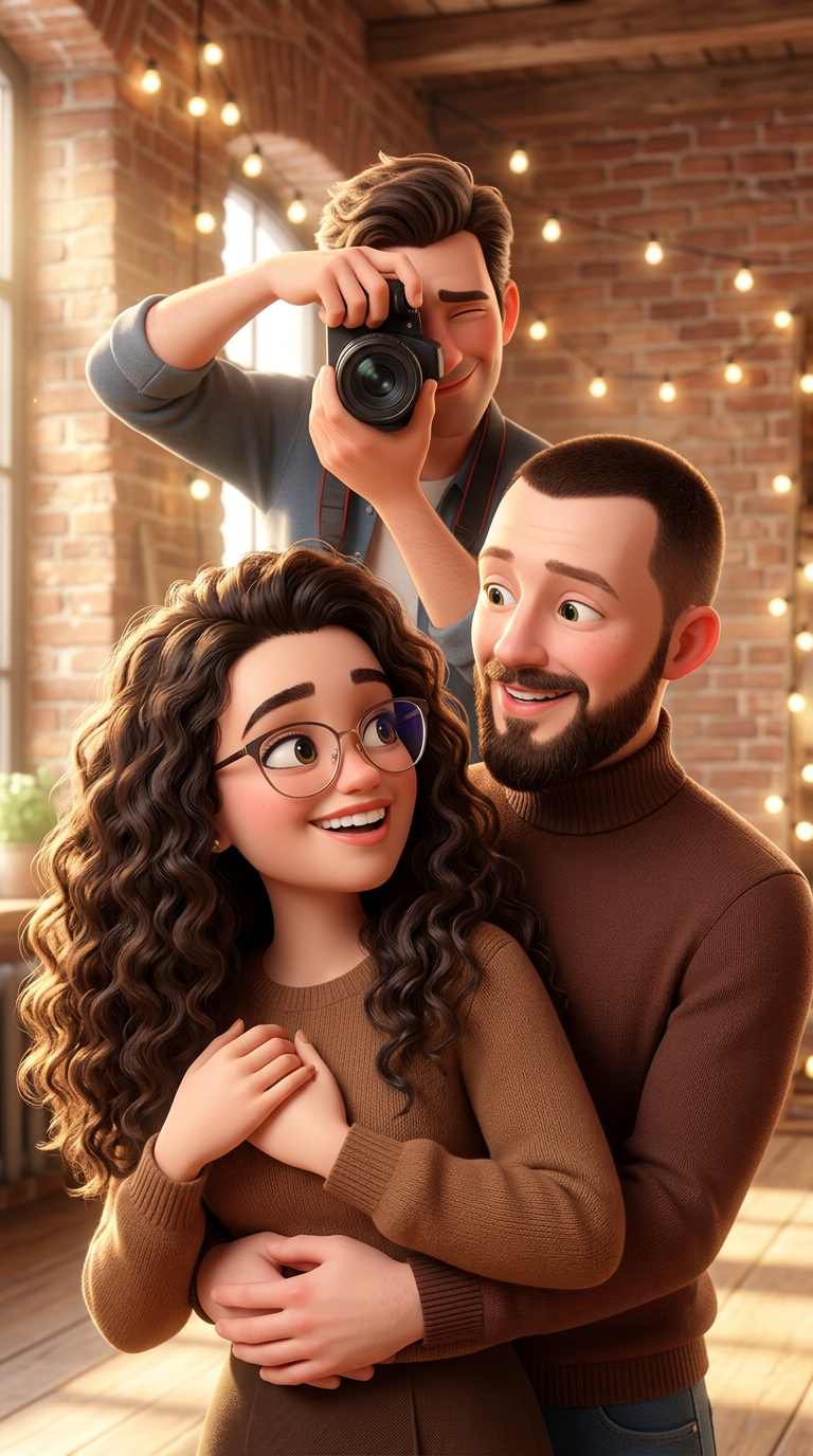
Сцена фотосессии в стиле "лав стори". .фото с референта + Профессиональный фотограф ловит момент.
━━━━━━━━━━━━━━━━...
 image
image
Art style: 3D Pixar.
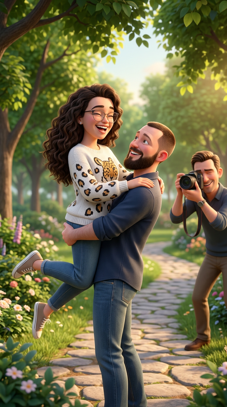
Сцена фотосессии в стиле "лав стори". Лика в белом свитере с принтом леопард.Павел кружит Лику на руках в красивом ...
 image
image
Art style: 3D Pixar.
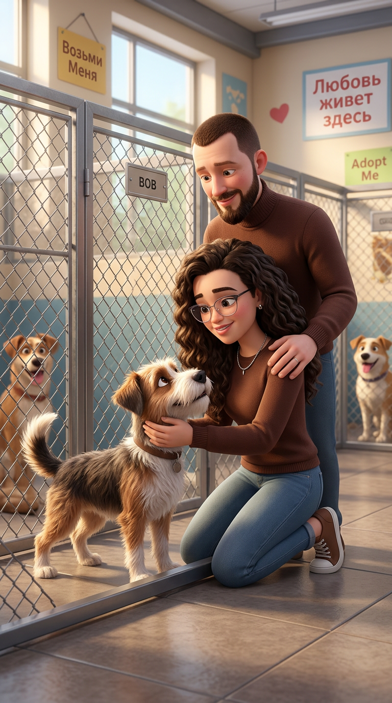
Пара в приюте для животных. Лика нежно гладит собаку в вольере, а Павел стоит рядом, обнимая ее за плечи. Вокруг др...
 image
image
Art style: 3D Pixar.
Зимний парк. Лика и Павел сидят на скамейке, укутанные в шарфы. Павел протягивает Лике коробку с аппетитными булочк...
 image
image
Art style: 3D Pixar.
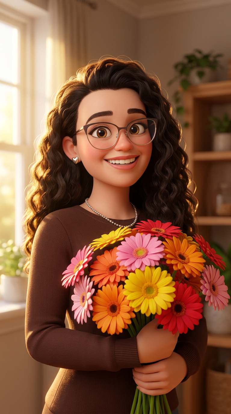
Лика держит в руках красивые разноцветные герберы. Утренний свет падает на цветы, создавая теплую и радостную атмос...
 image
image
Art style: 3D Pixar.
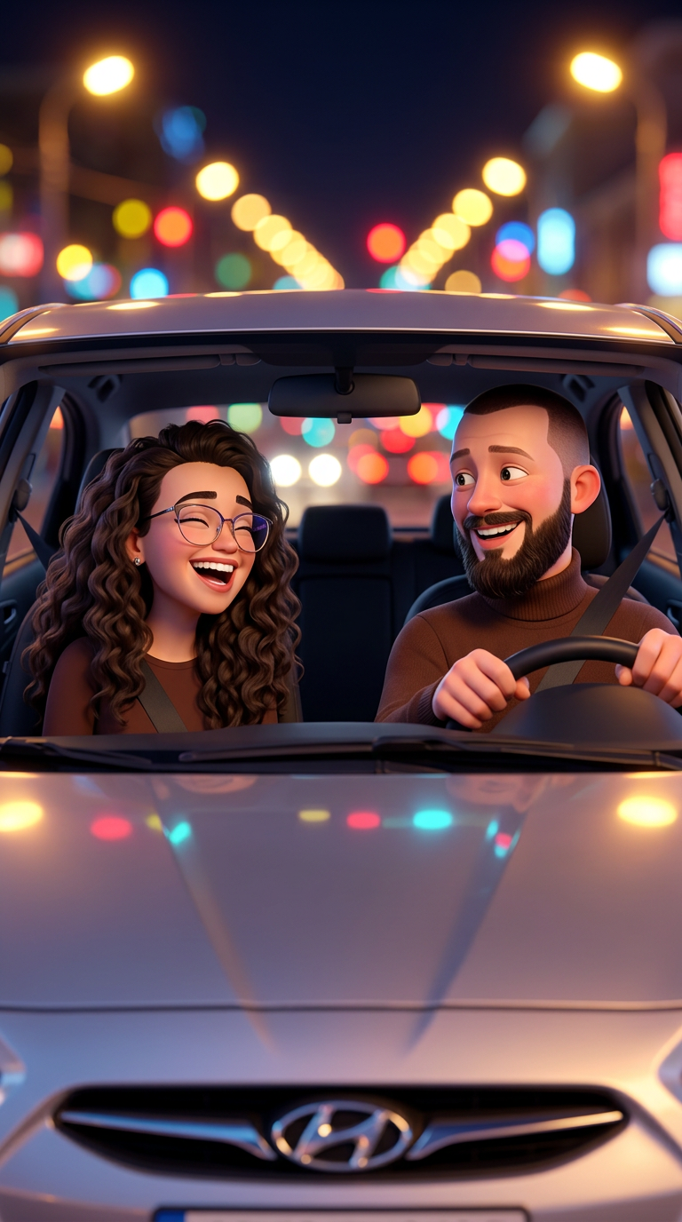
Павел и Лика в салоне серебристого Hyundai Solaris. Он за рулем, она на пассажирском сиденье. Они смеются, разговар...
 image
image
Art style: 3D Pixar.
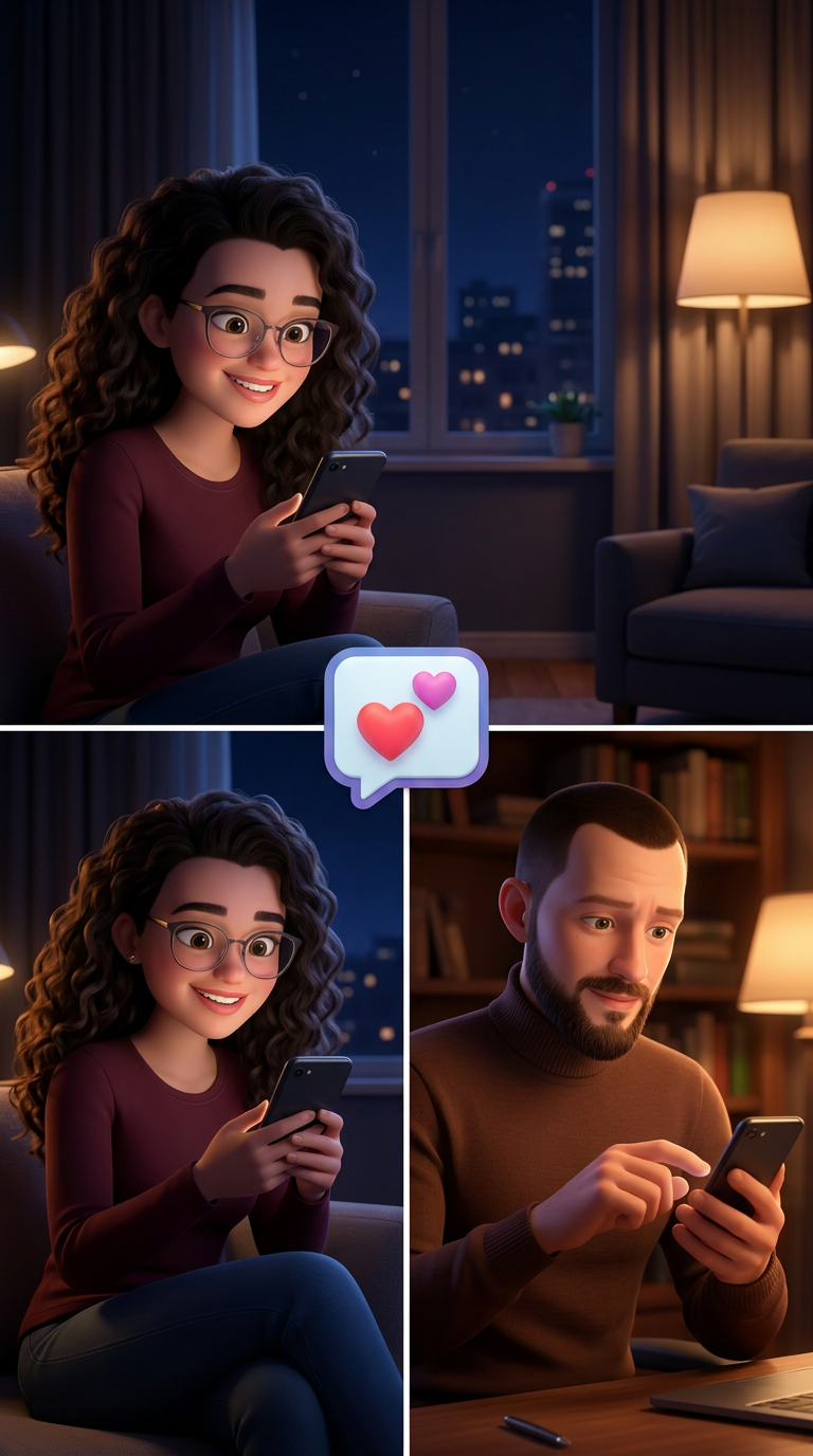
Коллаж из двух кадров: слева Лика с улыбкой смотрит в телефон, сидя в уютном кресле ночью. Справа Павел, также увле...
 image
image
Strong rule: style --- 3D Pixar ---. Красивая молодая женщина, около 28-30 лет, со светлыми волосами до плеч, выразительными карими глаза...
 image
image
154D6BE0-80FB-4FA8-AD0C-500B41334711.webp
 image
image
A1C0DE4A-29B2-4FD8-B19D-348803BA3F1E.webp
 image
image
Art style: Стиль 3 D.
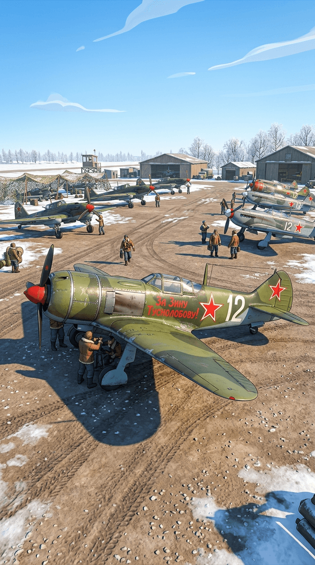
Советский истребитель времен ВОВ на аэродроме. На борту самолета яркой красной краской выведена надпись: 'За Зину ...

Плавный, медленный отъезд камеры (zoom out) от ее лица, чтобы показать всю фотографию. Эффект 'Ken Burns'.
On-screen dialogue (character...
 image
image
Art style: Реализм.
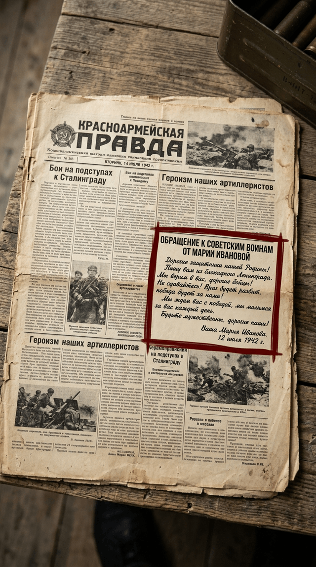
Крупный план разворота старой фронтовой газеты, пожелтевшей от времени. Выделена статья с ее обращением. Текст письм...
 image
image
Art style: Реализм.
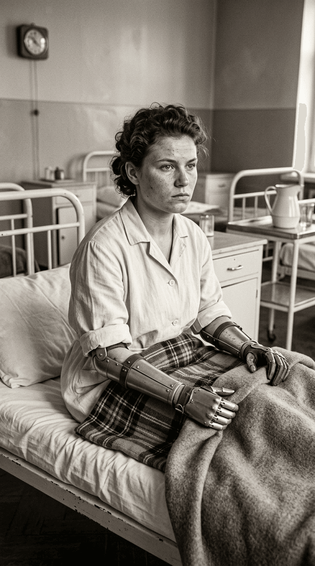
Реалистичное воссоздание фотографии: молодая Зинаида сидит на больничной койке, на ее лице видна несгибаемая воля. Е...
 image
image
Art style: Реализм.
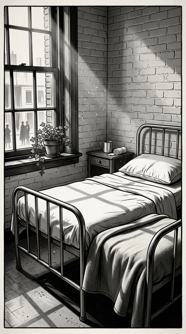
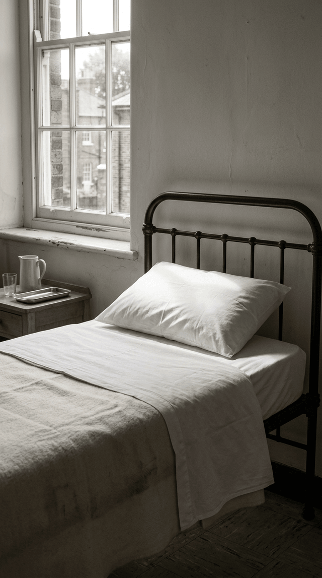
Больничная палата 1940-х годов. Стерильная, минималистичная обстановка. Крупный план аккуратно заправленной койки с ...
 image
image
Art style: Реализм.
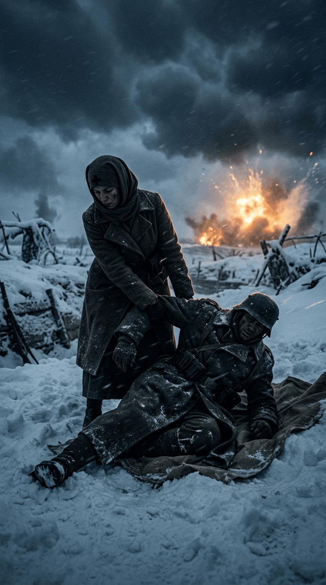
Художественная реконструкция. Темный силуэт женщины-медсестры тащит раненого солдата по глубокому снегу во время мет...
 image
image
Art style: Реализм.
Больничная палата 1940-х годов. Стерильная, минималистичная обстановка. Крупный план аккуратно заправленной койки с ...

Очень медленный, плавный наезд камеры (zoom in) на медаль, создавая ощущение важности и торжественности.
On-screen dialogue (characters ...
 image
image
Art style: Реализм.
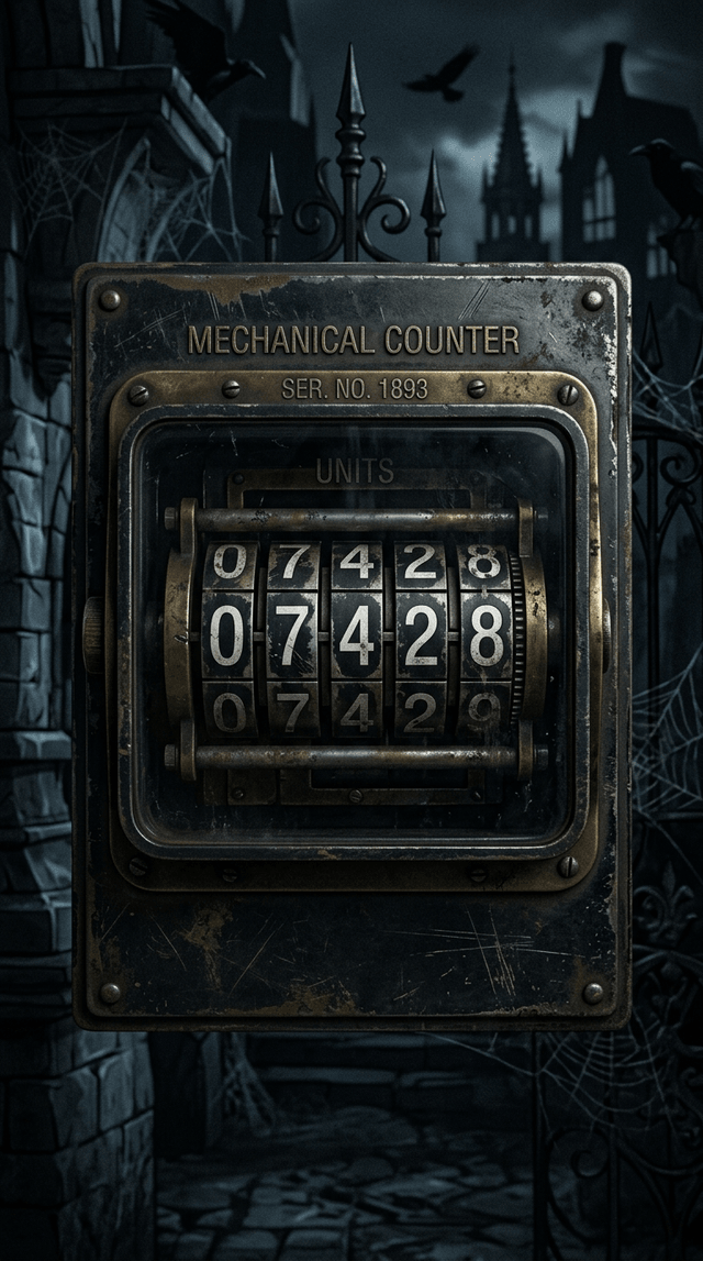
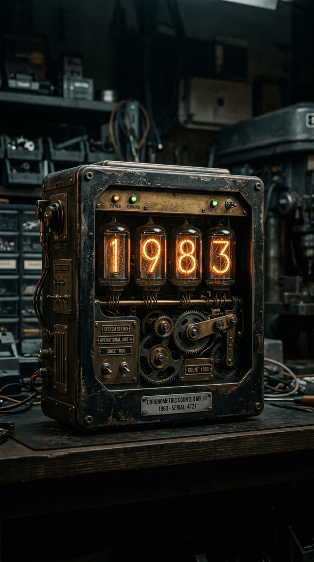
Стилизованная анимированная графика на темном фоне. В центре экрана большой цифровой счетчик, выполненный в стиле ст...
 image
image
Art style: Реализм.
Стилизованная анимированная графика на темном фоне. В центре экрана большой цифровой счетчик, выполненный в стиле ст...
 image
image
Art style: Реализм.
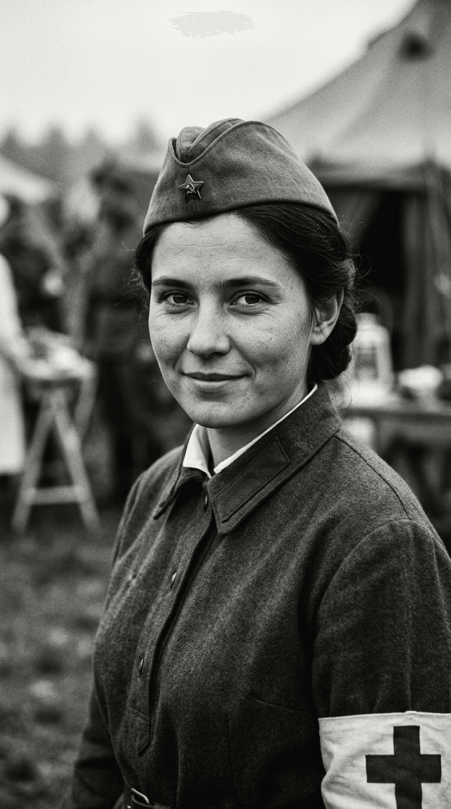
Атмосферная черно-белая фотография молодой красивой девушки (Зинаиды) в военной форме медсестры времен ВОВ. Она смот...
 image
image
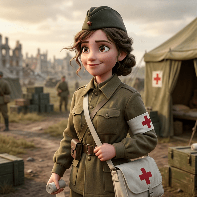
Strong rule: style --- Реализм ---. Strong rule: style --- 3D Pixar ---. Зинаида Туснолобова-Марченко, молодая красивая девушка 22 лет, с...
 image
image
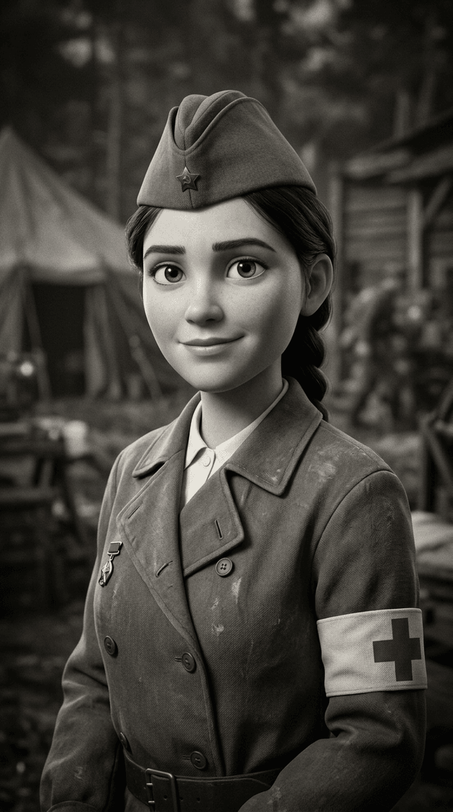
Атмосферная черно-белая фотография молодой красивой девушки (Зинаиды) в военной форме медсестры времен ВОВ. Она смотрит в камеру с мягкой...